Bone Analysis
The separately licensed Bone Analysis module for Dragonfly is a pre-clinical research application designed for the evaluation of high-resolution micro-CT image data of bone specimens. Bone Analysis provides 3D vector-based mappings of surface and volume anisotropy magnitude and directionality and 3D scalar-based mappings of volume fraction (see Mappings). In addition, an automated separation of cortical and trabecular bone in whole bone specimens drives the calculation of common bone morphometric indices that provide researchers with additional quantitative descriptions of bone micro-architecture (see Morphometric Indices).
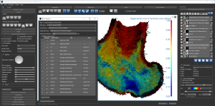
Bone Analysis dialog and Dragonfly workspace
3D vector-based mappings of anisotropy magnitude and orientation, as well as 3D scalar based mappings of volume fraction, can be computed for partial and whole bone specimens and then visualized and evaluated in 3D views. Measurements can be thresholded and different color maps can be applied to optimize visualizations (see Computing and Visualizing Mappings of Anisotropy and Volume Fraction).
Mappings of anisotropy and volume fraction
In the panel above, the image on the left shows anisotropy mapped by magnitude, with red being very anisotropic and blue isotropic. The image in the middle show the same map replotted in such a way that the color corresponds to orientation, while the image on the right shows the volume fraction of the segmented bone tissue.
Dragonfly's Bone Analysis module can separately identify and segment cortical and trabecular bone in whole-bone specimens and then compute common bone morphometric measurements, as described by Bouxsein et al*. Both global measurements and slice-by-slice measurements are available to evaluate bone micro-architecture (see Computing Cortical and Trabecular Measurements).
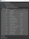
Global measurements of cortical and trabecular indices (see Computing Global Measurements) are available directly in the Bone Analysis dialog, shown below. Computed results can be exported in comma-separated values files (*.csv extension).
Bone Analysis dialog
In addition to the global measurements available for bone morphometric indices, per slice measurements in any oblique for segmented areas are available in the Slice Analysis dialog (see Computing Slice-by-Slice Measurements). The input for computing slice-by-slice measurements can be any region of interest — segmented bone, cortical bone, or trabeculae. Computed results can be exported in comma-separated values files (*.csv extension).
Slice Analysis dialog
Computing Cortical and Trabecular Measurements
Computing and Visualizing Mappings of Anisotropy and Volume Fraction


